

")


Polyostotic fibrous dysplasia: imaging findings of a controversial case
- Authors: De Michele F.1,2, Carpagnano F.A.1,2, Paparella M.T.1,2, Guglielmi G.1,2,3
-
Affiliations:
- Department of Clinical and Experimental Medicine, Foggia University School of Medicine
- Radiology Unit, Barletta University Campus UNIFG, “Dimiccoli” Hospital
- Radiology Unit, Hospital “Casa Sollievo Della Sofferenza”, San Giovanni Rotondo
- Issue: Vol 3, No 1 (2022)
- Pages: 55-63
- Section: Case reports
- URL: https://journal-vniispk.ru/DD/article/view/88605
- DOI: https://doi.org/10.17816/DD88605
- ID: 88605
Cite item

Abstract
Fibrous dysplasia is a rare non-neoplastic tumor-like congenital bone disease that is most likely associated with GNAS gene mutations, with a broad spectrum of clinical presentations, ranging from isolated monostotic and polyostotic forms to other extra-skeletal associated manifestations as in McCune–Albright syndrome. It is responsible for bone’s weakening and increased fragility, making it prone to fractures.
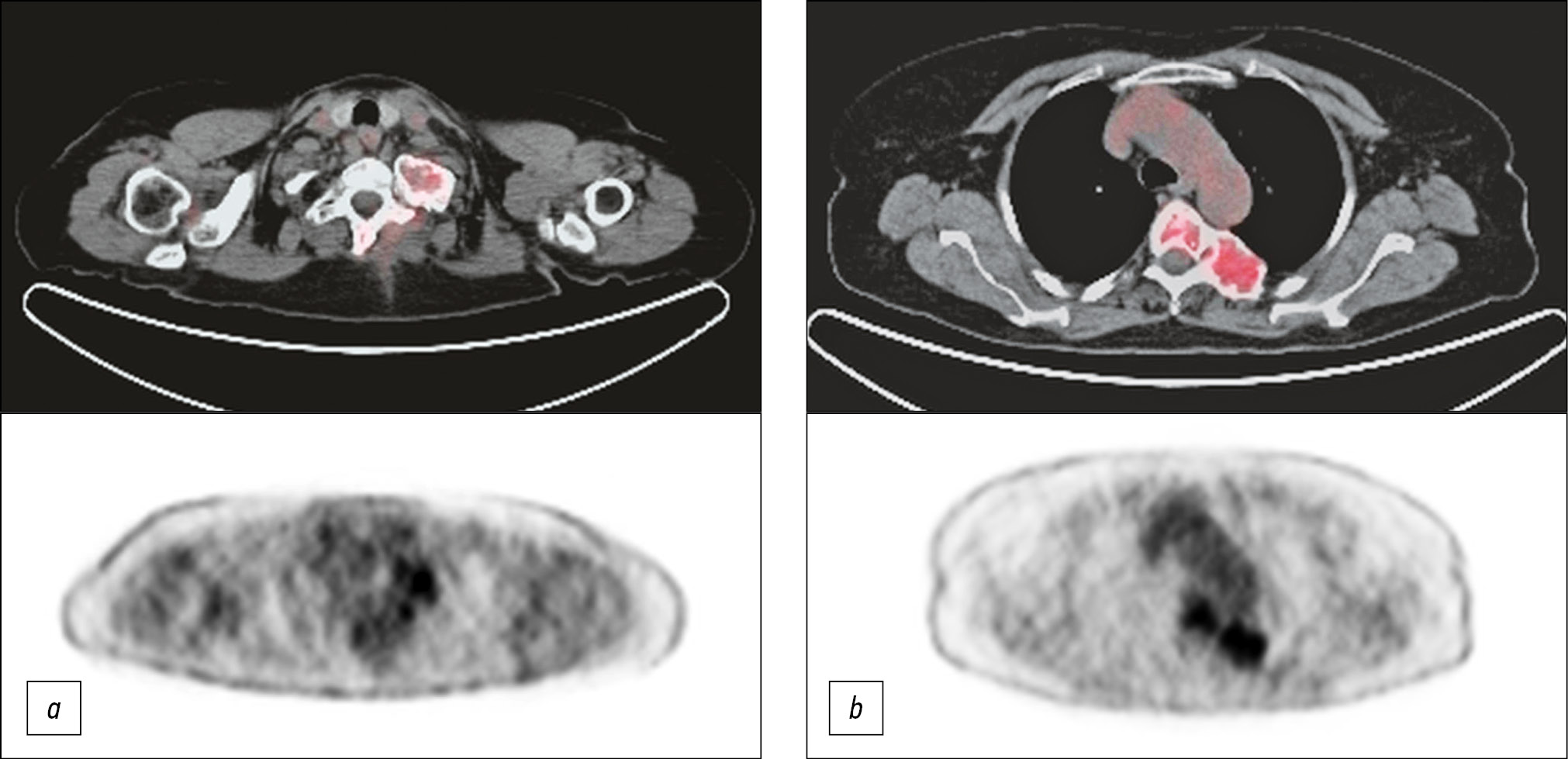
A 65-year-old female patient was referred to our radiology department for cervical and dorsal pain, with a previous diagnosis of incidental cervical and dorsal bone lesions that are suspected for metastases. X-ray, computed tomography, and magnetic resonance imaging were performed with a precise diagnostic suspicion of fibrous dysplasia that is confirmed by bone biopsy.
Fibrous dysplasia principally affects the bone and is characterized by bone replacement itself by dysplastic fibrous tissue. According to the number of affected bones and their association to endocrine alterations, it is classified into three categories monostotic, polyostotic, and Albright’s disease. Differential diagnosis with multiple myeloma among others and the best treatment decision was made.
Full Text
##article.viewOnOriginalSite##About the authors
Francesca De Michele
Department of Clinical and Experimental Medicine, Foggia University School of Medicine; Radiology Unit, Barletta University Campus UNIFG, “Dimiccoli” Hospital
Email: francesca.demichele82@gmail.com
ORCID iD: 0000-0002-6043-6362
Italy, Foggia; Foggia
Francesca A. Carpagnano
Department of Clinical and Experimental Medicine, Foggia University School of Medicine; Radiology Unit, Barletta University Campus UNIFG, “Dimiccoli” Hospital
Email: c.francesca1991@gmail.com
ORCID iD: 0000-0001-7681-2898
Italy, Foggia; Foggia
Maria T. Paparella
Department of Clinical and Experimental Medicine, Foggia University School of Medicine; Radiology Unit, Barletta University Campus UNIFG, “Dimiccoli” Hospital
Email: mt.paparella@gmail.com
ORCID iD: 0000-0003-2573-9509
Italy, Foggia; Foggia
Giuseppe Guglielmi
Department of Clinical and Experimental Medicine, Foggia University School of Medicine; Radiology Unit, Barletta University Campus UNIFG, “Dimiccoli” Hospital; Radiology Unit, Hospital “Casa Sollievo Della Sofferenza”, San Giovanni Rotondo
Author for correspondence.
Email: giuseppe.guglielmi@unifg.it
ORCID iD: 0000-0002-4325-8330
Medical Doctor, Full Professor of Radiology, Department of Clinical and Experimental Medicine.
Italy, Foggia; Foggia; FoggiaReferences
- Neville BW, Damm DD, Allen CM, Bouquot JE. Bone pathology. In: Neville BW, Damm DD, Allen CM, Bouquot JE, editors. Oral and maxillofacial pathology. Philadelphia, PA: WB Saunders Company; 2002. Р. 553–557.
- Iseri PK, Efendi H, Demirci A, Komsuoglu S. Fibrous dysplasia of the cranial bones: a case report and review of the literature. Yale J Biol Med. 2005;78(3):141–145.
- Lustig LR, Holliday MJ, McCarthy FF, Nager GT. Fibrous dysplasia involving the skull base and temporal bone. Arch Otolaryngol Head Neck Surg. 2001;127(10):1239–1247. doi: 10.1001/archotol.127.10.1239
- Chapurlat RD, Orcel P. Fibrous dysplasia of bone and McCune-Albright syndrome. Best Pract Res Clin Rheumatol. 2008;22(1):55–69. doi: 10.1016/j.berh.2007.11.004
- Feller L, Wood N, Khammissa R, et al. The nature of fibrous dysplasia. Head Face Med. 2009;5:22. doi: 10.1186/1746-160X-5-22
- Riminucci M, Kuznetsov SA, Cherman N, et al. Osteoclastogenesis in fibrous dysplasia of bone: in situ and in vitro analysis of IL-6 expression. Bone. 2003;33(3):434–442. doi: 10.1016/s8756-3282(03)00064-4
- Kransdorf MJ, Moser RP, Gilkey FW. Fibrous dysplasia. Radiographics. 1990;10(3):519–537. doi: 10.1148/radiographics.10.3.2188311
- Bhattacharyya N, Wiench M, Dumitrescu C, et al. Mechanism of FGF23 processing in fibrous dysplasia. J Bone Miner Res. 2012;27(5):1132–1141. doi: 10.1002/jbmr.1546
- Yamamoto T, Imanishi Y, Kinoshita E, et al. The role of fibroblast growth factor 23 for hypophosphatemia and abnormal regulation of vitamin D metabolism in patients with McCune-Albright syndrome. J Bone MinerMetab. 2005;23(3):231–237. doi: 10.1007/s00774-004-0589-9
Supplementary files


