

")


通过核磁共振纤维束成像、功能性核磁共振成像和脑电图分析中风后大脑下半球广泛损伤导致轻度神经功能缺损的临床病例。
- 作者: Gumin I.S.1, Gulyaev S.A.1, Beregov M.M.1, Lelyuk V.G.1
-
隶属关系:
- Federal center of brain research and neurotechnologies of the Federal Medical Biological Agency
- 期: 卷 5, 编号 3 (2024)
- 页面: 601-612
- 栏目: 临床病例及临床病例的系列
- URL: https://journal-vniispk.ru/DD/article/view/310041
- DOI: https://doi.org/10.17816/DD624967
- ID: 310041
如何引用文章
全文:

详细
包括大脑皮层在内的大脑不同部位受损后,神经功能缺损和生活质量下降的严重程度会有很大差异,通常与损伤量无关。病理变化的定位起着重要作用。 众所周知,优势半球和次优势半球的病变在临床表现和患者生活质量下降的程度上都有显著差异。
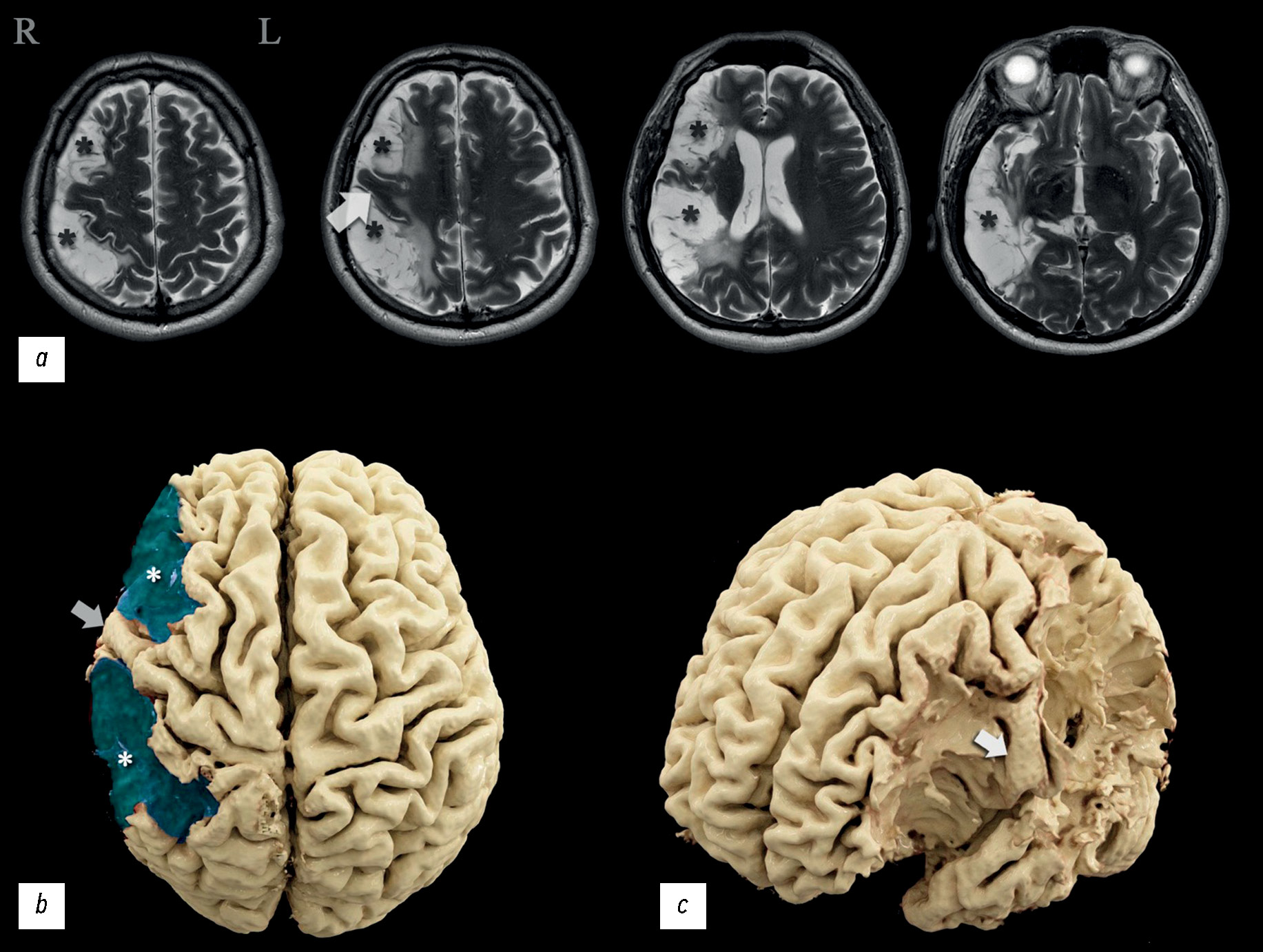
在临床病例分析中,一名患者在两次缺血性脑卒中后入院接受康复治疗,神经科医生和神经心理学家对其进行了检查,并通过脑电图、核磁共振成像、灌注评估计算机断层扫描、核磁共振纤维束成像和功能性核磁共振成像进行了全面的仪器检查。患者左侧肢体轻微瘫痪,自主活动调节能力障碍,神经动态指数轻度下降,注意力轻度下降,对自己的病情持批评态度。神经影像学检查结果发现,大脑中动脉区域的右侧次优势大脑半球存在广泛的梗塞后损伤。
显示脑损伤量与临床表现严重程度之间的失衡,并分析造成失衡的可能原因。根据功能研究的数据,确定优势半球,并提出功能中心重组的可能变体。与类似临床病例进行比较,分析其与本文的关系。所获得的信息扩展着运动执行、语言功能和运算能力主题改变区的认识。
作者简介
Ivan S. Gumin
Federal center of brain research and neurotechnologies of the Federal Medical Biological Agency
编辑信件的主要联系方式.
Email: ivangumin@mail.ru
ORCID iD: 0000-0003-2360-3261
SPIN 代码: 3454-2665
Scopus 作者 ID: 57223430019
俄罗斯联邦, Moscow
Sergey A. Gulyaev
Federal center of brain research and neurotechnologies of the Federal Medical Biological Agency
Email: gulyaev@fccps.ru
ORCID iD: 0000-0003-0549-0961
MD, Dr. Sci. (Medicine)
俄罗斯联邦, MoscowMikhail M. Beregov
Federal center of brain research and neurotechnologies of the Federal Medical Biological Agency
Email: mikhailberegov@gmail.com
ORCID iD: 0000-0003-1899-8131
SPIN 代码: 2559-0307
俄罗斯联邦, Moscow
Vladimir G. Lelyuk
Federal center of brain research and neurotechnologies of the Federal Medical Biological Agency
Email: vglelyuk@fccps.ru
ORCID iD: 0000-0002-9690-8325
MD, Dr. Sci. (Medicine), Professor
俄罗斯联邦, Moscow参考
- Global Health Estimates 2016: Deaths by cause, age, sex, by country and by region, 2000-2016 [Internet]. Geneva: World Health Organization; 2018 [cited 25.07.2019]. Available from: https://www.who.int/data/gho/data/themes/mortality-and-global-health-estimates
- Benjamin EJ, Muntner P, Alonso A, et al. Heart Disease and Stroke Statistics—2019 Update: A Report From the American Heart Association. Circulation. 2019;139(10):e56–e528. doi: 10.1161/CIR.0000000000000659
- Mijajlović MD, Pavlović A, Brainin M, et al. Post-stroke dementia — a comprehensive review. BMC Medicine. 2017;15(1):11. doi: 10.1186/s12916-017-0779-7
- Pohjasvaara T, Erkinjuntti T, Ylikoski R, et al. Clinical Determinants of Poststroke Dementia. Stroke. 1998;29(1):75–81. doi: 10.1161/01.str.29.1.75
- Barrett AM. Spatial Neglect and Anosognosia After Right Brain Stroke. Continuum (Minneapolis, Minn.). 2021;27(6):1624–1645. doi: 10.1212/CON.0000000000001076
- Broussolle E, Reynolds EH. Anglo-French neurological interactions in the 19th and early 20th centuries: Physicians, places and events. Revue neurologique. 2021;177(8):859–870. doi: 10.1016/j.neurol.2020.10.013
- Chakrabarty M, Pflieger EM, Cardillo E, Chatterjee A. Effects of Chronic Brain Injury on Quality of Life: A Study in Patients With Left- or Right-Sided Lesion. Archives of Rehabilitation Research and Clinical Translation. 2019;2(1):100031. doi: 10.1016/j.arrct.2019.100031
- Howard G, Till JS, Toole JF, et al. Factors Influencing Return to Work Following Cerebral Infarction. JAMA. 1985;253(2):226–232. doi: 10.1001/jama.1985.03350260078030
- Penfield W, Rasmussen T. The cerebral cortex of man. New York: Macmillan Company; 1950.
- Halligan P. Half a brain is enough: the story of Nico. Journal of Neurology Neurosurgery & Psychiatry. 2001;71(4):566. doi: 10.1136/jnnp.71.4.566b
- Gumin IS, Gubskiy IL, Mironov MB, et al. Dyke–Davidoff–Masson syndrome: description of clinical case with diagnostics by EEG, MRI, MR-tractography, fMRI. Neuromuscular Diseases. 2021;11(1):47–57. doi: 10.17650/2222-8721-2021-11-1-47-57
- Agris AR, Almazova AA, Altuhova TA, et al. Narusheniya pis’ma i chteniya u detej: izuchenie i korrekciya. Moscow: “LOGOMAG”; 2018. (In Russ.)
- Bain JS, Yeatman JD, Schurr R, et al. Evaluating arcuate fasciculus laterality measurements across dataset and tractography pipelines. Human Brain Mapping. 2019;40(13):3695–3711. doi: 10.1002/hbm.24626
- Roiha K, Kirveskari E, Kaste M, et al. Reorganization of the primary somatosensory cortex during stroke recovery. Clinical Neurophysiology. 2011;122(2):339–345. doi: 10.1016/j.clinph.2010.06.032
- Sanchez-Panchuelo RM, Francis S, Bowtell R, Schluppeck D. Mapping Human Somatosensory Cortex in Individual Subjects With 7T Functional MRI. Journal of neurophysiology. 2010;103(5):2544–2556. doi: 10.1152/jn.01017.2009
- Alary F, Doyon B, Loubinoux I, et al. Event-Related Potentials Elicited by Passive Movements in Humans: Characterization, Source Analysis, and Comparison to fMRI. Neuroimage. 1998;8(4):377–390. doi: 10.1006/nimg.1998.0377
- Cramer SC, Moore CI, Finklestein SP, Rosen BR. A Pilot Study of Somatotopic Mapping After Cortical Infarct. Stroke. 2000;31(3):668–671. doi: 10.1161/01.str.31.3.668
- Roux FE, Boulanouar K, Ibarrola D, et al. Functional MRI and intraoperative brain mapping to evaluate brain plasticity in patients with brain tumours and hemiparesis. Journal of Neurology Neurosurgery & Psychiatry. 2000;69(4):453–463. doi: 10.1136/jnnp.69.4.453
- Arsalidou M, Taylor MJ. Is 2+2=4? Meta-analyses of brain areas needed for numbers and calculations. Neuroimage. 2011;54(3):2382–2393. doi: 10.1016/j.neuroimage.2010.10.009
- Hawes Z, Sokolowski HM, Ononye CB, Ansari D. Neural underpinnings of numerical and spatial cognition: An fMRI meta-analysis of brain regions associated with symbolic number, arithmetic, and mental rotation. Neuroscience and Biobehavioral Reviews. 2019;103:316–336. doi: 10.1016/j.neubiorev.2019.05.007
- Fedorenko E, Blank IA. Broca’s Area Is Not a Natural Kind. Trends in Cognitive Sciences. 2020;24(4):270–284. doi: 10.1016/j.tics.2020.01.001
- Bach-y-Rita P. Brain plasticity as a basis for recovery of function in humans. Neuropsychologia. 1990;28(6):547–554. doi: 10.1016/0028-3932(90)90033-k
- Wan CY, Schlaug G. Music Making as a Tool for Promoting Brain Plasticity across the Life Span. Neuroscientist. 2010;16(5):566–577. doi: 10.1177/1073858410377805
- Nudo RJ. Remodeling of cortical motor representations after stroke: implications for recovery from brain damage. Molecular Psychiatry. 1997;2(3):188–191. doi: 10.1038/sj.mp.4000188
- Nudo RJ. Postinfarct Cortical Plasticity and Behavioral Recovery. Stroke. 2007;38(2):840–845. doi: 10.1161/01.STR.0000247943.12887.d2
补充文件


