


Analysis of long-term (34 years) observation of a patient with ophthalmological manifestations of Grenblad–Strandberg syndrome
- Authors: Sharma A.A.1, Zumbulidze N.G.1, Boyko E.V.1,2, Kononov A.V.1
-
Affiliations:
- North-Western State Medical University named after I.I. Mechnikov
- S.N. Fyodorov Eye Microsurgery Federal State Institution, Saint Petersburg Branch
- Issue: Vol 17, No 2 (2024)
- Pages: 81-93
- Section: Case reports
- URL: https://journal-vniispk.ru/ov/article/view/263005
- DOI: https://doi.org/10.17816/OV623597
- ID: 263005
Cite item

Abstract
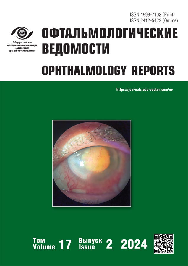
The prognosis for the life of patients with genetic pathology depends on the interaction between specialists from different areas of medicine for timely detection and selection of treatment tactics. Pseudoxanthoma elasticum (Grenblad–Strandberg syndrome) is a hereditary disease in which elastic fibers of the skin, the cardiovascular system and the retina are affected. Clinical manifestations: skin changes in Grenblad–Strandberg syndrome are represented by flat xanthomatous nodules of yellowish color. Cardiovascular manifestations of pseudoxanthoma elasticum are angina pectoris, decreased pulse amplitude, cardiomyopathy, sudden heart failure, often leading to death. Eye disorders occur in stages. For the early stages, the appearance of angioid streaks is typical, which appear as a result of calcification of elastic fibers of capillaries. The progression of the process leads to neovascularization and hemorrhages from the choriocapillaries, the formation of a subretinal neovascular membrane of foveolar localization, causing a decrease in vision. The late stages are characterized by scarring. Therapy depends on the stage and rate of progression of the disease and is effective at stages I–II (according to Vivaldi). Own clinical observation: Male patient, 71 years old, referred for cataract surgery with the diagnosis “Both eyes: Senile cataract, open-angle glaucoma, (stage I a, under beta-blocker therapy), Grenblad–Strandberg syndrome”. Attention is drawn to the long observation period — 34 years, with documented data from the first examinations in 1989 and all subsequent ones. Of particular interest is the availability of preserved patient documentation for all years of follow-up, including diagnosis and treatment.
Full Text
##article.viewOnOriginalSite##About the authors
Anton A. Sharma
North-Western State Medical University named after I.I. Mechnikov
Author for correspondence.
Email: saa98@mail.ru
ORCID iD: 0000-0003-4849-7004
SPIN-code: 1166-3917
Scopus Author ID: 58181373600
Russian Federation, Saint Petersburg
Nataliya G. Zumbulidze
North-Western State Medical University named after I.I. Mechnikov
Email: zumbulenok@mail.ru
ORCID iD: 0000-0002-7729-097X
SPIN-code: 4439-8855
MD, Cand. Sci. (Medicine), Assistant Professor
Russian Federation, Saint PetersburgErnest V. Boyko
North-Western State Medical University named after I.I. Mechnikov; S.N. Fyodorov Eye Microsurgery Federal State Institution, Saint Petersburg Branch
Email: Boiko111@list.ru
ORCID iD: 0000-0002-7413-7478
SPIN-code: 7589-2512
Scopus Author ID: 34067475000
MD, Dr. Sci. (Medicine), Professor
Russian Federation, Saint Petersburg; Saint PetersburgAnatoliy V. Kononov
North-Western State Medical University named after I.I. Mechnikov
Email: 7435020@gmail.com
ORCID iD: 0000-0002-4673-5024
Russian Federation, Saint Petersburg
References
- Conrath J, Matoni F. Angioid striae. Ophthalmologie. 2012;9(2):1–7. doi: 10.1016/j.jfo.2012.05.003 (In France).
- Timokhov VL, Rusanovskaya AV. Grönblad–Strandberg syndrome. Ophthalmology Reports. 2014;7(4):69–72. EDN: THSCLH
- Kryazheva SS, Snarskaya ES, Kartashova MG, Filatova IV. Grönblad–Strandberg syndrome. Russian Journal of Skin and Venereal Diseases. 2011;(5):43–47. EDN: OHXPYV
- Fukumoto T, Iwanaga A, Fukunaga A, et al. First-genetic analysis of atypical phenotype of pseudoxanthoma elasticum with ocular manifestations in the absence of characteristic skin lesions. J Eur Acad Dermatol Venereol. 2018;32(4):e147–e149. doi: 10.1111/jdv.14637
- Jiang Q, Endo M, Dibra F, et al. Pseudoxanthoma elasticum is a metabolic disease. J Invest Dermatol. 2009;129(2):348–354. doi: 10.1038/jid.2008.212
- Orkin VF, Platonova AN, Marchenko VM. Pseudoxanthoma elasticum (Grönblad–Strandberg syndrome). Russian Journal of Clinical Dermatology and Venereology. 2008;3(6):44–46. EDN: QIZBOZ
- Wolff K, Goldsmith L, Katz S, et al. Fitzpatrick’s dermatology in general medicine. 8th ed. (2 Volume Set). McGraw-Hill; 2012. P. 1426–1429.
- Rigal D. Observation for use in the history of xanthelmic diffuse chéloide. Annals of dermatology and syphiligraphy. 1881;2: 491–495. (In France).
- Knapp H. On the formation of dark angioid streaks as an unusual metamorphosis of retinal hemorrage. Arch Ophthalmol. 1892;21:289–292.
- Finger RP, Charbel Issa P, Ladewig MS, et al. Pseudoxanthoma elasticum: genetics, clinical manifestations and therapeutic approaches. Surv Ophthalmol. 2009;54(2):272–285. doi: 10.1016/j.survophthal.2008.12.006
- Groenblad E. Angioid streaks — Pseudoxanthoma elasticum; vorläufig Mitteilung. Acta Ophthalmol. 1929;7:329
- Jensen OA. Bruchs membrane in pseudoxanthoma elasticum. Histochemical, ultrastructural, and X-ray microanalytical study of the membrane and angioid streak areas. Albrecht Von Graefes Arch Klin Exp Ophthalmol. 1977;203(3–4):311–320. doi: 10.1007/BF00409836
- Clarkson JG, Altman RD. Angioid streaks. Surv Ophthalmol. 1982;26(5):235–246. doi: 10.1016/0039-6257(82)90158-8
- Jampol LM, Acheson R, Eagle RC, et al. Calcification of Bruch’s membrane in angioid streaks with homozygous sickle cell disease. Arch Ophthalmol. 1987;105(1):93–98. doi: 10.1001/archopht.1987.01060010099039
- Baccarani-Contri M, Vincenzi D, Cicchetti F, et al. Immunochemical identification of abnormal constituents in the dermis of pseudoxanthoma elasticum patients. Eur J Histochem. 1994;38(2):111–113.
- Quaglino D, Boraldi F, Barbieri D, et al. Abnormal phenotype of in vitro dermal fibroblasts from patients with Pseudoxanthoma elasticum (PXE). Biochim Biophys Acta. 2000;1501(1):51–62. doi: 10.1016/s0925-4439(00)00007-7
- Gotting C, Sollberg S, Kuhn J, et al. Serum xylosyltransferase: a new biochemical marker of the sclerotic process in systemic sclerosis. J Invest Dermatol. 1999;112(6):919–924. doi: 10.1046/j.1523-1747.1999.00590.x
- Uitto J. Pseudoxanthoma elasticuma connective tissue disease or a metabolic disorder at the genome/environment interface? J Invest Dermatol. 2004;122:9–10. doi: 10.1111/j.0022-202X.2004.22350.x
- Aessopos A, Farmakis D, Loukopoulos D. Elastic tissue abnormalities resembling pseudoxanthoma elasticum in beta thalassemia and the sickling syndromes. Blood. 2002;99:30–35. doi: 10.1182/blood.v99.1.30
- Pasquali-Ronchetti I, Garcia-Fernandez MI, Boraldi F, et al. Oxidative stress in fibroblasts Ofrom patients with pseudoxanthoma elasticum: possible role in the pathogenesis of clinical manifestations. J Pathol. 2006;208(1):54–61. doi: 10.1002/path.1867
- Bergen AA, Plomp AS, Schuurman EJ, et al. Mutations in ABCC6 cause pseudoxanthoma elasticum. Nat Genet. 2000;25(2):228–231. doi: 10.1038/76109
- Le Saux O, Urban Z, Tschuch C, et al. Mutations in a gene encoding an ABC transporter cause pseudoxanthoma elasticum. Nat Genet. 2000;25(2):223–227. doi: 10.1038/76102
- Ringpfeil F, Lebwohl MG, Uitto J. Abstracts: mutations in the MRP6 gene cause pseudoxanthoma elasticum. J Invest Dermatol. 2000;115(2):332. doi: 10.1046/j.1523-1747.2000.00abs.x
- Gorgels TG, Hu X, Scheffer GL, et al. Disruption of ABCC6 in the mouse: novel insight in the pathogenesis of pseudoxanthoma elasticum. Hum Mol Genet. 2005;14(13):1763–1773. doi: 10.1093/hmg/ddi183
- Chassaing N, Martin L, Mazereeuw J, et al. Novel ABCC6 mutations in pseudoxanthoma elasticum. J Invest Dermatol. 2004;122(3):608–613. doi: 10.1111/j.0022-202X.2004.22312.x
- Gheduzzi D, Guidetti R, Anzivino C, et al. ABCC6 mutations in Italian families affected by pseudoxanthoma elasticum (PXE). Hum Mutat. 2004;24(5):438–439. doi: 10.1002/humu.9284
- Le Saux O, Beck K, Sachsinger C, et al. A spectrum of ABCC6 mutations is responsible for pseudoxanthoma elasticum. Am J Hum Genet. 2001;69(4):749–764. doi: 10.1086/323704
- Aessopos A, Farmakis D, Loukopoulos D. Elastic tissue abnormalities in inherited haemolytic syndromes. Eur J Clin Invest. 2002;32(9):640–642. doi: 10.1046/j.1365-2362.2002.01033.x
- Elouarradi H, Abdelouahed K. Angioid streaks. Pan Afr Med J. 2014;17:13. doi: 10.11604/pamj.2014.17.13.3609
- Challenor VF, Conway N, Monro JL. The surgical treatment of restrictive cardiomyopathy in pseudoxanthoma elasticum. Br Heart J. 1988;59(2):266–269. doi: 10.1136/hrt.59.2.266
- Fukuda K, Uno K, Fujii T, et al. Mitral stenosis in pseudoxanthoma elasticum. Chest. 1992;101(6):1706–1707. doi: 10.1378/chest.101.6.1706
- Lebwohl M, Halperin J, Phelps RG. Brief report: occult pseudoxanthoma elasticum in patients with premature cardiovascular disease. N Engl J Med. 1993;329(17):1237–1239. doi: 10.1056/NEJM199310213291705
- Krill AE, Klien BA, Archer DB. Precursors of angioid streaks. Am J Ophthalmol. 1973;76(6):875–879. doi: 10.1016/0002-9394(73)90076-7
- Gills JP Jr, Paton D. Mottled fundus oculi in pseudoxanthoma elasticum; a report on two siblings. Arch Ophthalmol. 1965;73: 792–795. doi: 10.1001/archopht.1965.00970030794007
- Benitez-Herreros J, Camara-Gonzalez C, Lopez-Guajardo L, et al. Choroidal neovascularization secondary to angioid streaks: A familial case report. Arch Soc Esp Oftalmol. 2014;89(5):190–193. doi: 10.1016/j.oftal.2012.11.005 (In Spanish).
- Mansour AM, Ansari NH, Shields JA, et al. Evolution of angioid streaks. Ophthalmologica. 1993;207(2):57–61. doi: 10.1159/000310407
- Groenblad E. Color photographs of angioid streaks in the late stages. Acta Ophthalmol (Copenh). 1958;36(3):472–474.
- Carlborg U, Ejrup B, Groenblad E, Lund F. Vascular studies in pseudoxanthoma elasticum and angioid streaks; with a series of color photographs of the eyeground lesions. Acta Med Scand Suppl. 1959;350:1–84.
- Connor PJ Jr, Juergens JL. Pseudoxanthoma elasticum and angioid streaks. A review of 106 cases. Am J Med. 1961;30:537–543. doi: 10.1016/0002-9343(61)90078-x
- Secretan M, Zografos L, Guggisberg D, Piguet B. Chorioretinal vascular abnormalities associated with angioid streaks and pseudoxanthoma elasticum. Arch Ophthalmol. 1998;116(10):1333–1336. doi: 10.1001/archopht.116.10.1333
- Shields JA, Federman JL, Tomer TL, et al. Angioid streaks. I. Ophthalmoscopic variations and diagnostic problems. Br J Ophthalmol. 1975;59(5):257–266. doi: 10.1136/bjo.59.5.257
- Gelisken O, Hendrikse F, Deutman AF. A long-term follow-up study of laser coagulation of neovascular membranes in angioid streaks. Am J Ophthalmol. 1988;105(3):299–303. doi: 10.1016/0002-9394(88)90014-1
- Fine HF. Photodynamic therapy in the anti-VEGF era. Br J Ophthalmol. 2007;91(6):707–708. doi: 10.1136/bjo.2007.114041
- Brown DM, Regillo CD. Anti-VEGF agents in the treatment of neovascular age-related macular degeneration: applying clinical trial results to the treatment of everyday patients. Am J Ophthalmol. 2007;144(4):627–637. doi: 10.1016/j.ajo.2007.06.039
- Pieramici DJ, Rabena MD. Anti-VEGF therapy: comparison of current and future agents. Eye (Lond). 2008;22(10):1330–1336. doi: 10.1038/eye.2008.88
- Ahmadieh H, Taei R, Soheilian M, et al. Single-session photodynamic therapy combined with intravitreal bevacizumab for neovascular age-related macular degeneration. Eur J Ophthalmol. 2008;18(2):297–300. doi: 10.1177/112067210801800222
- Georgalas I, Papaconstantinou D, Koutsandrea C, et al. Angioid streaks, clinical course, complications, and current therapeutic management. Ther Clin Risk Manag. 2009;5(1):81–89. doi: 10.2147/TCRM.S4682
- Machemer R, Steinhorst UH. Retinal separation, retinotomy, and macular relocation: II. A surgical approach for age-related macular degeneration? Graefes Arch Clin Exp Ophthalmol. 1993;231(11): 635–641. doi: 10.1007/BF00921957
- Roth DB, Estafanous M, Lewis H. Macular translocation for subfoveal choroidal neovascularization in angioid streaks. Am J Ophthalmol. 2001;131(3):390–392. doi: 10.1016/s0002-9394(99)00207-x
- Toth CA, Freedman SF. Macular translocation with 360-degree peripheral retinectomy impact of technique and surgical experience on visual outcomes. Retina. 2001;21(4):293–303. doi: 10.1097/00006982-200108000-00001
- Toth CA, Lapolice DJ, Banks AD, Stinnett SS. Improvement in near visual function after macular translocation surgery with 360-degree peripheral retinectomy. Graefes Arch Clin Exp Ophthalmol. 2004;242(7):541–548. doi: 10.1007/s00417-004-0867-1
- Park CH, Toth CA. Macular translocation surgery with 360-degree peripheral retinectomy following ocular photodynamic therapy of choroidal neovascularization. Am J Ophthalmol. 2003;136(5): 830–835. doi: 10.1016/s0002-9394(03)00723-2
- de Juan E Jr, Fujii GY. Limited macular translocation. Eye. 2001;15:413–423. doi: 10.1038/eye.2001.146
- Fujii GY, Humayun MS, Pieramici DJ, et al. Initial experience of inferior limited macular translocation for subfoveal choroidal neovascularization resulting from causes other than age-related macular degeneration. Am J Ophthalmol. 2001;131(1):90–100. doi: 10.1016/s0002-9394(00)00769-8
- Tanaka M, Shimada H, Haruyama M, et al. Surgical removal of choroidal neovascularization in angioid streaks. Nippon Ganka Gakkai Zasshi. 2003;107(8):440–444.
Supplementary files


