

")


Experience with artificial intelligence algorithms for the diagnosis of vertebral compression fractures based on computed tomography: from testing to practical evaluation
- Authors: Artyukova Z.R.1, Petraikin A.V.1, Kudryavtsev N.D.1, Petryaykin F.A.2, Semenov D.S.1, Sharova D.E.1, Belaya Z.E.3, Vladzimirskyy A.V.1,4, Vasilev Y.A.1
-
Affiliations:
- Research and Practical Clinical Center for Diagnostics and Telemedicine Technologies
- Lomonosov Moscow State University
- Endocrinology Research Centre
- The First Sechenov Moscow State Medical University
- Issue: Vol 5, No 3 (2024)
- Pages: 505-518
- Section: Original Study Articles
- URL: https://journal-vniispk.ru/DD/article/view/310034
- DOI: https://doi.org/10.17816/DD624250
- ID: 310034
Cite item
Full Text

Abstract
BACKGROUND: Osteoporosis is often diagnosed at the stage with complications, i.e., low-energy fractures. Vertebral compression fractures, which are complications of osteoporosis and predictors of subsequent fractures, are often asymptomatic. Compression fractures can be found by computed tomography performed for other indications with vertebral morphometry. Approaches to using artificial intelligence algorithms designed for diagnosing vertebral compression fractures were analyzed.
AIM: Testing artificial intelligence algorithms to conduct morphometric analysis of vertebrae on chest computed tomography scans and assess the possibility of their implementation in medical organizations of the Moscow Healthcare Department.
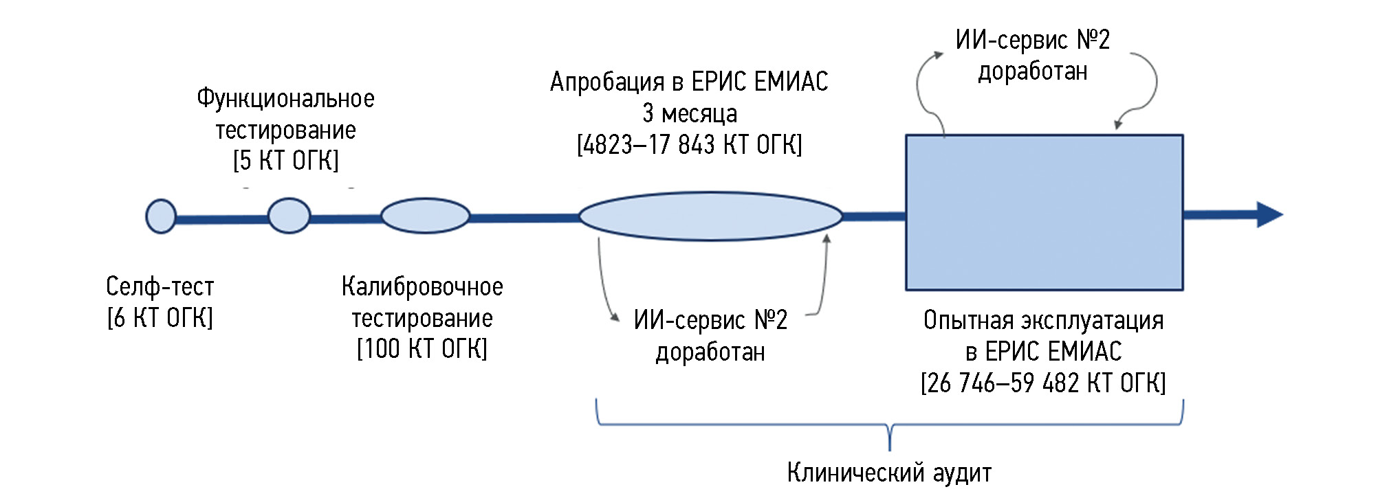
MATERIALS AND METHODS: To set a clinical task for artificial intelligence algorithms, basic diagnostic requirements in the area of “vertebral compression fractures (osteoporosis)” were formulated. The testing of the artificial intelligence algorithms included the following stages: self-testing, functional and calibration testing, practical evaluation, and operation testing. The first three stages of testing were performed using previously generated datasets. At practical evaluation and operation testing, artificial intelligence algorithms analyzed the data from computed tomography performed in medical organizations. The expert group of radiologists assessed the diagnostic accuracy and functional capacity of the AI algorithms at all stages. The resulting quantitative metrics of the accuracy of artificial intelligence algorithms were compared with the required target values.
RESULTS: From June 2021 to June 2022, two artificial intelligence algorithms (Nos. 1 and 2) with different methods of detecting compression fractures were tested. Both artificial intelligence algorithms successfully passed the self-testing (6 tests), functional (5 tests), and calibration (100 tests) stages. The area under the ROC curve for artificial intelligence algorithm No. 1 was 0.99 (95% CI, 0.98–1), and for artificial intelligence algorithm No. 2, it was 0.91 (95% CI, 0.85–0.96). Artificial intelligence algorithm No. 1 passed the practical evaluation stage without any significant remarks, whereas algorithm No. 2 was sent for fine-tuning. After the operation testing stage, the following accuracy metrics were obtained: the areas under the ROC curve for artificial intelligence algorithm Nos. 1 and 2 were 0.93 (95% CI, 0.89–0.96) and 0.92 (95% CI, 0.90–0.94), respectively. At all stages, both artificial intelligence algorithms demonstrated sufficient metrics for clinical validation.
CONCLUSION: Artificial intelligence algorithms for the automatic diagnosis of vertebral compression fractures have been tested, demonstrating the high quality of their operation. artificial intelligence algorithms can be applied as a supplementary tool in the medical decision support system.
Full Text
##article.viewOnOriginalSite##About the authors
Zlata R. Artyukova
Research and Practical Clinical Center for Diagnostics and Telemedicine Technologies
Author for correspondence.
Email: zl.artyukova@gmail.com
ORCID iD: 0000-0003-2960-9787
SPIN-code: 7550-2441
Russian Federation, Moscow
Alexey V. Petraikin
Research and Practical Clinical Center for Diagnostics and Telemedicine Technologies
Email: alexeypetraikin@gmail.com
ORCID iD: 0000-0003-1694-4682
SPIN-code: 6193-1656
MD, Dr. Sci. (Medicine), Assistant Professor
Russian Federation, MoscowNikita D. Kudryavtsev
Research and Practical Clinical Center for Diagnostics and Telemedicine Technologies
Email: KudryavtsevND@zdrav.mos.ru
ORCID iD: 0000-0003-4203-0630
SPIN-code: 1125-8637
Russian Federation, Moscow
Fedor A. Petryaykin
Lomonosov Moscow State University
Email: feda.petraykin@gmail.com
ORCID iD: 0000-0001-6923-3839
SPIN-code: 7803-1005
Russian Federation, Moscow
Dmitry S. Semenov
Research and Practical Clinical Center for Diagnostics and Telemedicine Technologies
Email: semenovds4@zdrav.mos.ru
ORCID iD: 0000-0002-4293-2514
SPIN-code: 2278-7290
Cand. Sci. (Engineering)
Russian Federation, MoscowDaria E. Sharova
Research and Practical Clinical Center for Diagnostics and Telemedicine Technologies
Email: SharovaDE@zdrav.mos.ru
ORCID iD: 0000-0001-5792-3912
SPIN-code: 1811-7595
Russian Federation, Moscow
Zhanna E. Belaya
Endocrinology Research Centre
Email: jannabelaya@gmail.com
ORCID iD: 0000-0002-6674-6441
SPIN-code: 4746-7173
MD, Dr. Sci. (Medicine)
Russian Federation, MoscowAnton V. Vladzimirskyy
Research and Practical Clinical Center for Diagnostics and Telemedicine Technologies; The First Sechenov Moscow State Medical University
Email: VladzimirskijAV@zdrav.mos.ru
ORCID iD: 0000-0002-2990-7736
SPIN-code: 3602-7120
MD, Dr. Sci. (Medicine)
Russian Federation, Moscow; MoscowYuriy A. Vasilev
Research and Practical Clinical Center for Diagnostics and Telemedicine Technologies
Email: VasilevYA1@zdrav.mos.ru
ORCID iD: 0000-0002-0208-5218
SPIN-code: 4458-5608
MD, Cand. Sci. (Medicine)
Russian Federation, MoscowReferences
- Belaya ZhE, Belova KYu, Biryukova EV, et al. Federal clinical guidelines for diagnosis, treatment and prevention of osteoporosis. Osteoporosis and Bone Diseases. 2021;24(2):4–47. doi: 10.14341/osteo12930
- Petraikin A, Artyukova Z, Nisovtsova LA, et al. Analysis of the effectiveness of implementing screening of osteoporosis. Manager Zdravoochranenia. 2021;2:31–39. doi: 10.21045/1811-0185-2021-2-31-39
- Alacreu E, Moratal D, Arana E. Opportunistic screening for osteoporosis by routine CT in Southern Europe. Osteoporosis International. 2017;28(3):983–990. doi: 10.1007/s00198-016-3804-3
- Ziemlewicz TJ, Binkley N, Pickhardt PJ. Opportunistic Osteoporosis Screening: Addition of Quantitative CT Bone Mineral Density Evaluation to CT Colonography. Journal of the American College of Radiology. 2015;12(10):1036–1041. doi: 10.1016/j.jacr.2015.04.018
- Rebello D, Anjelly D, Grand DJ, et al. Opportunistic screening for bone disease using abdominal CT scans obtained for other reasons in newly diagnosed IBD patients. Osteoporosis international. 2018;29(6):1359–1366. doi: 10.1007/s00198-018-4444-6
- Artyukova ZR, Kudryavtsev ND, Petraikin AV, et al. Using an artificial intelligence algorithm to assess the bone mineral density of the vertebral bodies based on computed tomography data. Medical Visualization. 2023;27(2):125–137. doi: 10.24835/1607-0763-1257
- Jang S, Graffy PM, Ziemlewicz TJ, et al. Opportunistic osteoporosis screening at routine abdominal and Thoracic CT: Normative L1 trabecular attenuation values in more than 20 000 adults. Radiology. 2019;291(2):360–367. doi: 10.1148/radiol.2019181648
- Smets J, Shevroja E, Hügle T, et al. Machine Learning Solutions for Osteoporosis-A Review. J Bone Miner Res. 2021;36(5):833–851. doi: 10.1002/jbmr.4292
- Petraikin AV, Skripnikova IA. Quantitative Computed Tomography, modern data. Review. Medical Visualization. 2021;25(4):134–146. doi: 10.24835/1607-0763-1049
- Lenchik L, Rogers LF, Delmas PD, et al. Diagnosis of Osteoporotic Vertebral Fractures: Importance of Recognition and Description by Radiologists. American Journal of Roentgenology. 2004;183(4):949–958. doi: 10.2214/ajr.183.4.1830949
- Pinto A, Berritto D, Russo A, et al. Traumatic fractures in adults: Missed diagnosis on plain radiographs in the Emergency Department. Acta Biomedica. 2018;89:111–123. doi: 10.23750/abm.v89i1-S.7015
- Carberry GA, Pooler BD, Binkley N, et al. Unreported vertebral body compression fractures at abdominal multidetector CT. Radiology. 2013;268(1):120–126. doi: 10.1148/radiol.13121632
- Vladzimirskii AV, Vasil’ev YuA, Arzamasov KM, et al. Computer Vision in Radiologic Diagnostics: The First Stage of the Moscow Experiment: Monograph. 2nd edition, revised and supplemented. Moscow: Izdatel’skie resheniya; 2023. (In Russ.) EDN: FOYLXK
- Genant HK, Wu CY, Cornelis van K, et al. Vertebral fracture assessment using a semiquantitative technique. Journal of Bone and Mineral Research. 1993;8(9):1137–1148. doi: 10.1002/jbmr.5650080915
- Mosmed.ai [Internet]. State Budgetary Institution of Healthcare of the City of Moscow “Scientific and Practical Clinical Center for Diagnostics and Telemedicine Technologies of the Department of Healthcare of the City of Moscow” [cited 2024 Mar 14]. (In Russ.)Available from: https://mosmed.ai/
- Clinical guidelines. Osteoporosis. [Internet]. Ministry of Health of the Russian Federation. [cited 2023 Oct 24]. Available from: https://cr.minzdrav.gov.ru/schema/87_4
- The Adult Official Positions of the ISCD [Internet]. The International Society For Clinical Densitometry [cited 2023 Oct 24]. Available from: https://iscd.org/official-positions-2023/
- ACR–SPR–SSR practice parameter for the performance of quantitative computed tomography (QCT) bone mineral density [Internet]. American College of Radiology [cited 2023 Oct 24]. Available from: https://www.acr.org/-/media/ACR/Files/Practice-Parameters/qct.pdf
- Certificate of the Russian Federation on state registration of the database № 2023621171/ 11.04.2023. Vasil’ev YuA, Turavilova EV, Vladzimirskii AV, et al. MosMedData: CT scan with signs of osteoporosis of the spine. Available from: https://www.elibrary.ru/download/elibrary_52123357_73775308.PDF [cited 2023 Oct 23]. (In Russ.) EDN: SHLWTC
- Pisov M, Kondratenko V, Zakharov A, et al. Keypoints Localization for Joint Vertebra Detection and Fracture Severity Quantification. In: Martel AL, et al. Medical Image Computing and Computer Assisted Intervention – MICCAI 2020. MICCAI 2020. Lecture Notes in Computer Science. Vol. 12266. Springer; 2020. P:723–732. doi: 10.1007/978-3-030-59725-2_70
- Bar A, Wolf BL, Orna A, et al. Compression fractures detection on CT. Medical Imaging 2017: Computer-Aided Diagnosis. 2017;10134:1013440. doi: 10.48550/arXiv.1706.01671
- Lesnyak O, Baranova I, Belova K, et al. Osteoporosis in Russian Federation: epidemiology, socio-medical and economical aspects (review). Traumatology and Orthopedics of Russia. 2018;24(1):155–168. doi: 10.21823/2311-2905-2018-24-1-155-168
- Seo JW, Lim SH, Jeong JG, et al. A deep learning algorithm for automated measurement of vertebral body compression from X-ray images. Sci Rep. 2021;11(1):13732. doi: 10.1038/s41598-021-93017-x
- Murata K, Endo K, Aihara T, et al. Artificial intelligence for the detection of vertebral fractures on plain spinal radiography. Sci Rep. 2020;10(1):20031. doi: 10.1038/s41598-020-76866-w
- Dong Q, Luo G, Lane NE, et al. Deep Learning Classification of Spinal Osteoporotic Compression Fractures on Radiographs using an Adaptation of the Genant Semiquantitative Criteria. Acad Radiol. 2022;29(12):1819–1832. doi: 10.1016/j.acra.2022.02.020
- Tomita N, Cheung YY, Hassanpour S. Deep neural networks for automatic detection of osteoporotic vertebral fractures on CT scans. Computers in Biology and Medicine. 2018;98:8–15. doi: 1016/j.compbiomed.2018.05.011
- Valentinitsch A, Trebeschi S, Kaesmacher J, et al. Opportunistic osteoporosis screening in multi-detector CT images via local classification of textures. Osteoporosis International. 2019;30(6):1275–1285. doi: 10.1007/s00198-019-04910-1
- Yasaka K, Akai H, Kunimatsu A, et al. Prediction of bone mineral density from computed tomography: application of deep learning with a convolutional neural network. Eur Radiol. 2020;30(6):3549–3557. doi: 10.1007/s00330-020-06677-0
- Nam KH, Seo I, Kim DH, et al. Machine Learning Model to Predict Osteoporotic Spine with Hounsfield Units on Lumbar Computed Tomography. J Korean Neurosurg Soc. 2019;62(4):442–449. doi: 10.3340/jkns.2018.0178
- Zhang J, Liu F, Xu J, et al. Qingqing. Automated detection and classification of acute vertebral body fractures using a convolutional neural network on computed tomography. Frontiers in Endocrinology. 2023;14(1132725):1–10. doi: 10.3389/fendo.2023.1132725
- Pickhardt PJ, Dustin PB, Travisи L, et al. Opportunistic Screening for Osteoporosis Using Abdominal Computed Tomography Scans Obtained for Other Indications. Annals of internal medicine. 2013;158(8):588. doi: 10.7326/0003-4819-158-8-201304160-00003
- Del Lama RS, Candido RM, Chiari-Correia NS, et al. Computer-Aided Diagnosis of Vertebral Compression Fractures Using Convolutional Neural Networks and Radiomics. J Digit Imaging. 2022;35(3):446–458. doi: 10.1007/s10278-022-00586-y
- Morozov SP, Gavrilov AV, Arkhipov IV, et al. Effect of artificial intelligence technologies on the CT scan interpreting time in COVID-19 patients in inpatient setting. Russian Journal of Preventive Medicine. 2022;25(1):14–20. doi: 10.17116/profmed20222501114
- Vladzymyrskyy AV, Kudryavtsev ND, Kozhikhina DD, et al. Effectiveness of using artificial intelligence technologies for dual descriptions of the results of preventive lung examinations. Russian Journal of Preventive Medicine. 2022;25(7):7–15. doi: 10.17116/profmed2022250717
- Shelepa AA, Petraikin AV, Artyukova ZR, et al. Artificial intelligence for bone mineral density assessment: general population data. Digital Diagnostics. 2022;3(S1):23–24. doi: 10.17816/DD10571
Supplementary files


